Home
/ Anatomy Of The Ribs And Chest : Figure 3 from Relevant surgical anatomy of the chest wall ... : Chest bone, ribs, lung, heart, xiphoid process, sternum anatomy.
Anatomy Of The Ribs And Chest : Figure 3 from Relevant surgical anatomy of the chest wall ... : Chest bone, ribs, lung, heart, xiphoid process, sternum anatomy.
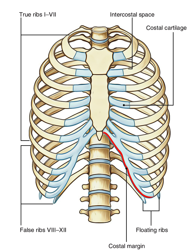
Anatomy Of The Ribs And Chest : Figure 3 from Relevant surgical anatomy of the chest wall ... : Chest bone, ribs, lung, heart, xiphoid process, sternum anatomy.. It plays important roles in the support of the spinal cord, ribcage, and muscles of the chest. It is made up of 12 pairs of ribs. 1).7within the anterior thorax, the first seven pairs of ribs are attached to the sternum, the 8th through 10th ribs are attached to each other by costal cartilage, and the 11th and 12th ribs remain unattached, or floating. The cartilage strips are called costal cartilage (costal is the anatomical adjective that refers to the rib) and connect on their other end to the sternum. The flail segment moves paradoxically during respiration, reducing the efficacy of ventilation.
Multiple rib fractures resulting in a flail chest alters the normal physiology of the thoracic wall; There are twelve pairs of ribs, all of which articulate with the vertebral column. The diaphragm is the piston. All ribs articulate posteriorly with the transverse processes and vertebral bodies of their respective thoracic vertebrae and the vertebral body directly superior (figure 1). Rib cage, in vertebrate anatomy, basketlike skeletal structure that forms the chest, or thorax, and is made up of the ribs and their corresponding attachments to the sternum (breastbone) and the vertebral column.
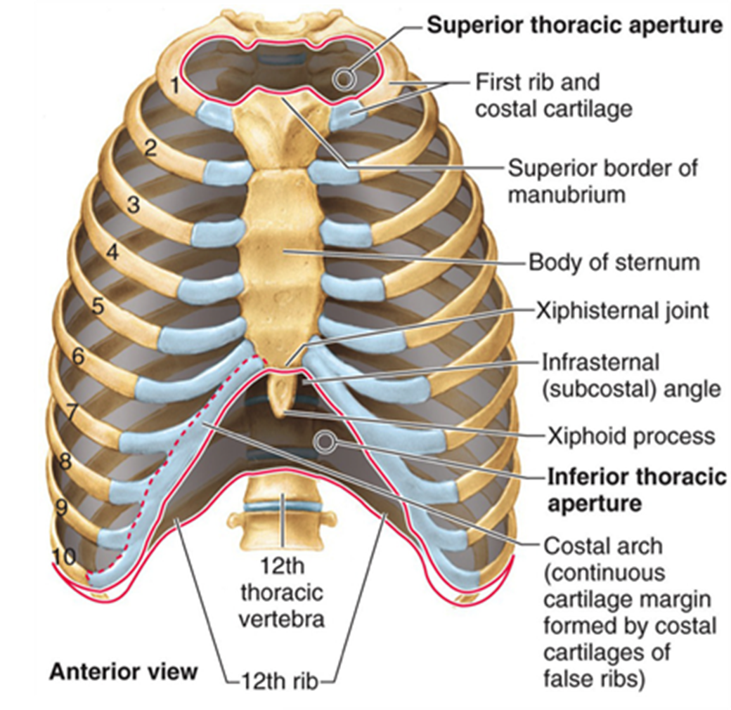
Easy Notes On 【Ribs】Learn in Just 4 Minutes! - Earth's Lab from www.earthslab.com Ten of the twelve ribs connect to strips of hyaline cartilage on the anterior side of the body. The mechanism of fracture can damage the intercostal muscles, vessels, and nerves resulting in weakness, hemorrhage, and muscle paralysis, respectively. The ribs, costal cartilages, and sternum form the firm supporting structure of the cylinder's wall. A part of the chest will then move independently from the rest of the chest wall. Ninja nerds!join us in this video where we show the sternum and rib articulation anatomy through the use of a model. It is made up of the manubrium superiorly, the body and the xiphisternum (figure 1).the manubrium has an upper central depression, the suprasternal notch. Structures to identify • heart • lungs • mediastinum • pleural space • chest wall • …everything else! The posterior rib (right) is farther from the film and is magnified more than the anterior rib (left), which is in contact with the film.
The cartilage strips are called costal cartilage (costal is the anatomical adjective that refers to the rib) and connect on their other end to the sternum.
First i'll do an intro to the different organs and structures in the chest, and then i'll go over some images showing their locations. It is made up of the manubrium superiorly, the body and the xiphisternum (figure 1).the manubrium has an upper central depression, the suprasternal notch. The ribs are curved, flat bones which form the majority of the thoracic cage. Ribs can also be divided into true, false and floating ribs: As part of the bony thorax, the ribs protect the internal thoracic organs. Contributing to their role in protecting the internal thoracic organs. The chest wall is formed from the sternum anteriorly, 12 pairs of ribs, costal cartilages and intercostal muscles laterally, and the thoracic vertebrae posteriorly. The human thorax includes the thoracic cavity and the thoracic wall. The bones of the chest and upper back combine to form the strong, protective rib cage around the vital thoracic organs such as the heart and lungs. Chest bone, ribs, lung, heart, xiphoid process, sternum anatomy. The remaining ribs are typical. In this image, you will find common carotid arteries, internal jugular vein, subclavian artery, subclavian vein, heart, right lung, 6th rib, diaphragm, costal cartilage in it. In insects, crustaceans, and the extinct trilobites, the thorax is one of the three main divisions of the creature's body, each of which is in turn composed of multiple segments.
It plays important roles in the support of the spinal cord, ribcage, and muscles of the chest. Flail chest describes what happens when blunt force trauma to the chest causes multiple adjacent ribs to fracture and separate from the chest wall. All ribs articulate posteriorly with the transverse processes and vertebral bodies of their respective thoracic vertebrae and the vertebral body directly superior (figure 1). The first step in understanding thorax anatomy is to find out its boundaries. The degree of rotation is best assessed by comparing the length of the anterior ribs visible on both sides.
MBBS DOCTORS: Basics of Reading Chest X ray | Nurse ... from i.pinimg.com Contributing to their role in protecting the internal thoracic organs. Anatomy of the chest cavity and sternum: However, only seven have a direct articulation with the sternum. All ribs articulate posteriorly with the transverse processes and vertebral bodies of their respective thoracic vertebrae and the vertebral body directly superior (figure 1). Each pair is numbered based on their attachment to the sternum, a bony process at the front of the rib cage which serves as an anchor point. The first step in understanding thorax anatomy is to find out its boundaries. The palpable midline sternum is variable in size and shape; Usually, the thorax is wider in transverse dimension than in the anteroposterior dimension.
They are extremely light, but highly resilient;
The diaphragm is the piston. The chest wall is formed from the sternum anteriorly, 12 pairs of ribs, costal cartilages and intercostal muscles laterally, and the thoracic vertebrae posteriorly. The mechanism of fracture can damage the intercostal muscles, vessels, and nerves resulting in weakness, hemorrhage, and muscle paralysis, respectively. The anatomy of the ribs and the sternum and their relationship to chest wall structure and function as with all parts of the body, the anatomy and physiology of the chest wall are intimately intertwined. Physiology of the chest wall if one were to look at the mechanics of respiration in a simplified manner, the easiest model to express the mechanics of respiration is a cylinder with a piston. The thoracic cage (rib cage) forms the thorax (chest) portion of the body. It consists of the 12 pairs of ribs with their costal cartilages and the sternum (figure 1). Chest bone, ribs, lung, heart, xiphoid process, sternum anatomy. These sections consist of the manubrium, body or corpus and the xiphoid process. It is made up of the manubrium superiorly, the body and the xiphisternum (figure 1).the manubrium has an upper central depression, the suprasternal notch. Usually, the thorax is wider in transverse dimension than in the anteroposterior dimension. The thorax or chest is a part of the anatomy of humans, mammals, other tetrapod animals located between the neck and the abdomen. The anatomy of the human ribs is made up of 24 ribs which are parted in 12 pairs (each on the left and right side of the chest wall), with the sternum, metasternum(the xiphoid process), and the costal cartilages all situated at the anterior of the chest wall, followed by the thoracic vertebrae on the posterior of the chest wall.
To carry out the unique functions performed by the chest wall, the anatomic structures are formed precisely for maximal efficiency. You will also find the xiphoid process, 10th rib, the apex of the heart, the coronary vein of the heart. Organs & structures of the chest heart. As newborn chest radiographs are taken in the ap plane, the. The ribs, costal cartilages, and sternum form the firm supporting structure of the cylinder's wall.
Thoracic, Chest & Rib Pain | Aligned for Life from alignedforlife.co.uk A part of the chest will then move independently from the rest of the chest wall. However, only seven have a direct articulation with the sternum. The rib cage also anchors the bones of the head, neck, shoulders, and arms to the trunk of the body. Moving during chest expansion to enable lung inflation. The sternum is made of three sections of bone tissue. Pectus excavatum is a congenital deformity of the ribs and the sternum producing a concave appearance of the anterior chest wall. The palpable midline sternum is variable in size and shape; These sections consist of the manubrium, body or corpus and the xiphoid process.
It is made up of 12 pairs of ribs.
Each are symmetrically paired on a right and left side. Multiple rib fractures resulting in a flail chest alters the normal physiology of the thoracic wall; Usually, the thorax is wider in transverse dimension than in the anteroposterior dimension. The bones of the chest and upper back combine to form the strong, protective rib cage around the vital thoracic organs such as the heart and lungs. A part of the chest will then move independently from the rest of the chest wall. In this image, you will find common carotid arteries, internal jugular vein, subclavian artery, subclavian vein, heart, right lung, 6th rib, diaphragm, costal cartilage in it. Physiology of the chest wall if one were to look at the mechanics of respiration in a simplified manner, the easiest model to express the mechanics of respiration is a cylinder with a piston. The rib cage is a bony structure found in the chest (thoracic cavity). Contributing to their role in protecting the internal thoracic organs. The anatomy of the ribs and the sternum and their relationship to chest wall structure and function as with all parts of the body, the anatomy and physiology of the chest wall are intimately intertwined. Moving during chest expansion to enable lung inflation. In insects, crustaceans, and the extinct trilobites, the thorax is one of the three main divisions of the creature's body, each of which is in turn composed of multiple segments. 1).7within the anterior thorax, the first seven pairs of ribs are attached to the sternum, the 8th through 10th ribs are attached to each other by costal cartilage, and the 11th and 12th ribs remain unattached, or floating.
The mechanism of fracture can damage the intercostal muscles, vessels, and nerves resulting in weakness, hemorrhage, and muscle paralysis, respectively anatomy of ribs. The diaphragm is the piston.